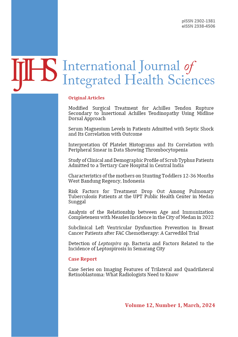
Case Series on Imaging Features of Trilateral and Quadrilateral Retinoblastoma: What Radiologists Need to Know
DOI:
https://doi.org/10.15850/ijihs.v12.n1.3359Keywords:
Pineal Gland, Quadrilateral, Retinoblastoma, Suprasellar, TrilateralAbstract
Objective: To identify the differences between trilateral and quadrilateral retinoblastoma based on radiologic imaging.
Case: We found three cases of retinoblastoma at Sanglah General Hospital. The retinoblastoma patients we found were boys under the age of 5. The first patient presented with swelling, pain, and impaired vision in the right eye. The second and third patients presented with eye protrusion and leukocoria. Computed tomography (CT) imaging revealed a calcified mass in the intraorbital region that extended to the cerebral hemispheres. It may be accompanied by hair-on-end periosteal reaction. In the first patient, the mass extended to the pineal gland. In the second and third patients, the mass extended to the suprasellar and pineal regions. Trilateral retinoblastoma consists of a primordial midline neuroectodermal tumor that originates in the pineal or suprasellar region. It is an uncommon combination of unilateral or bilateral retinoblastomas. Quadrilateral retinoblastoma, on the other hand, presents with bilateral retinoblastoma and tumors in both the suprasellar and pineal regions. The histopathology of trilateral and quadrilateral retinoblastoma shows a different pattern from retinoblastoma.
Conclusion: Retinoblastoma poses one of the most challenging problems due to its distinct patterns of growth, extension, and recurrence. Intracranial metastasis of trilateral and quadrilateral retinoblastoma exhibits a different pattern. The prognosis and diagnosis of retinoblastoma are significantly influenced by clinical examination, radiologic imaging, and histopathology.
References
- Silvera VM, Guerin JB, Brinjikji W, Dalvin LA. Retinoblastoma: what the neuroradiologist needs to know. AJNR Am J Neuroradiol. 2021;42(4):618–26. doi:10.3174/ajnr.A6949
- Gui T, Zheng H, Liu M, Xia Z, Ji X, Yin Q, et al. Clinical and magnetic resonance imaging features of 14 patients with trilateral retinoblastoma. Quant Imaging Med Surg. 2021;11(4):1458–69. doi:10.21037/qims-20-605
- Ali MJ, Honavar SG, Reddy VA. Distant metastatic retinoblastoma without central nervous system involvement. Indian J Ophthalmol. 2013;61(7):357–9. doi:10.4103/0301-4738.97077
- Jones J, Iqbal S, Worsley C, et al. Retinoblastoma. Radiopaedia. 2023 https://doi.org/10.53347/rID-1976
- Lim YS, Juraida E, Alagaratnam J, Menon BS. Trilateral retinoblastoma. Med J Malaysia. 2015;66(2):156–7. https://doi.org/10.3928/0191-3913-19830301-09
- Hisbulloh H, Hendara F. Serial case: unilateral dan trilateral retinoblastoma. Medica Hosp J Clin Med. 2020;7(1):119–29. https://doi.org/10.36408/mhjcm.v7i1.438
- Razek AAKA, Elkhamary S. MRI of retinoblastoma. Br J Radiol. 2014;84(1005):773–e190. https://doi.org/10.1259/bjr/32022497
- Habib YS, Youssef AA, Alkiki HA, Ghareeb HT, El H, Elzomor DA. High Resolution MR Imaging guidelines in retinoblastoma : prospective study correlated with histopathological results. Egypt J Radiol Nucl Med. 2020;3(51)1–11. https://doi.org/10.1186/s43055-020-0143-3
- Chen X, Wang J, Cao Z, Hosaka K, Jensen L, Yang H, et al. Invasiveness and metastasis of retinoblastoma in an orthotopic zebrafish tumor model. Sci Rep. 2015;5:10351. doi:10.1038/srep10351
- Sharma S, Kamala R, Nair D, et al. Round cell tumors: classification and immunohistochemistry. Indian J Med Paediatr Oncol. 2017;38(3):349–53. doi:10.4103/ijmpo.ijmpo_84_16
Downloads
Published
Issue
Section
License
Authors retain copyright and grant International Journal of Integrated Health Sciences right of first publication with the work simultaneously licensed under a Creative Commons Attribution License that allows others to freely share and remix the work with an acknowledgement of the work's authorship and initial publication in International Journal of Integrated Health Sciences.